Single Service
CT Scan
Service Information
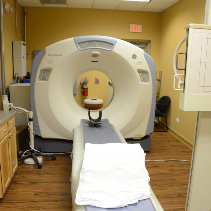
CT Scan
CT scanning—sometimes called CAT scanning—is a noninvasive medical test that helps physicians diagnose and treat medical conditions. It's one of the best and fastest tools for examining the chest, abdomen and pelvis because it provides detailed, cross-sectional views of all types of tissue.
Our technicians who operate the scanner are certified will stay in constant communication with you throughout your scan.
CT scans of internal organs, bones, soft tissue and blood vessels provide greater clarity and reveal more details than regular x-ray exams.
Some of Your Questions
- What is CT Scanning?
- How should I prepare?
- How is the CAT scan performed?
- What will I experience during and after the procedure?
- Who interprets the results and how do I get them?
- What is CT Scanning?
-
CT imaging is:
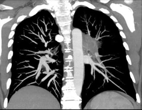
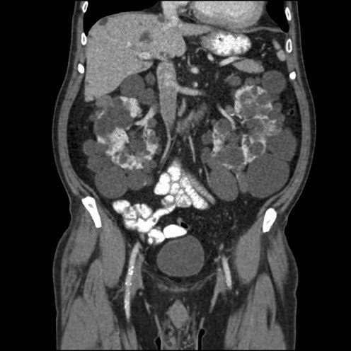
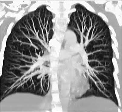
- one of the best and fastest tools for examining the chest, abdomen and pelvis because it provides detailed, cross-sectional views of all types of tissue.
- used to examine patients with severe injuries from incidents such as a motor vehicle accident.
- performed on patients with acute symptoms such as abdominal pain or difficulty breathing.
- often the best method for detecting many different cancers, including lung, liver, kidney and pancreatic cancer, since the image allows a physician to confirm the presence of a tumor and measure its size, precise location and the extent of the tumor's involvement with other nearby tissue.
- an examination that plays a significant role in the detection, diagnosis and treatment of vascular diseases that can lead to stroke, kidney failure or even death. CT is commonly used to assess for pulmonary embolism (a blood clot in the lung vessels) as well as for abdominal aortic aneurysms (AAA).
- invaluable in diagnosing and treating spinal problems and injuries to the hands, feet and other skeletal structures because it can clearly show even very small bones as well as surrounding tissues such as muscle and blood vessels.
In pediatric patients, CT is rarely used to diagnose tumors of the lung or pancreas as well as abdominal aortic aneurysms. For children, CT imaging is more often used to evaluate:
- lymphoma
- neuroblastoma
- kidney tumors
- congenital malformations of the heart, kidneys and blood vessels
- cystic fibrosis
- complications of acute appendicitis
- complications of pneumonia
- inflammatory bowel disease
- severe injuries
Physicians often use the CT examination to:
- quickly identify injuries to the lungs, heart and vessels, liver, spleen, kidneys, bowel or other internal organs in cases of trauma.
- guide biopsies and other procedures such as abscess drainages and minimally invasive tumor treatments.
- plan for and assess the results of surgery, such as organ transplants or gastric bypass.
- stage, plan and properly administer radiation treatments for tumors as well as monitor response to chemotherapy.
- measure bone mineral density for the detection of osteoporosis.
- How should I prepare?
-
You should wear comfortable, loose-fitting clothing to your exam. You may be given a gown to wear during the procedure.
Metal objects, including jewelry, eyeglasses, dentures and hairpins, may affect the CT images and should be left at home or removed prior to your exam. You may also be asked to remove hearing aids and removable dental work. Women will be asked to remove bras containing metal underwire. You may be asked to remove any piercings, if possible.
You should inform the technologist if you have a pacemaker. Pacemakers do not hinder the use of CT as in MRI as long as the scanner will not be taking images repeatedly over the area of the pacemaker device in the upper chest. This is usually not an issue for cardiac CT exams.
You may be asked not to eat or drink anything for a few hours beforehand, especially if a contrast material will be used in your exam. You should inform your physician of all medications you are taking and if you have any allergies. If you have a known allergy to contrast material, or "dye," your doctor may prescribe medications (usually a steroid) to reduce the risk of an allergic reaction. These medications generally need to be taken 12 hours prior to administration of contrast material. To avoid unnecessary delays, contact your doctor before the exact time of your exam.
Also inform your doctor of any recent illnesses or other medical conditions and whether you have a history of heart disease, asthma, diabetes, kidney disease or thyroid problems. Any of these conditions may increase the risk of an unusual adverse effect.
Women should always inform their physician and the CT technologist if there is any possibility that they may be pregnant. - How is the CAT scan performed?
-
The technologist begins by positioning you on the CT examination table, usually lying flat on your back or less commonly, on your side or on your stomach. Straps and pillows may be used to help you maintain the correct position and to help you remain still during the exam. Depending on the part of the body being scanned, you may be asked to raise your arms over your head.
Many scanners are fast enough that children can be scanned without sedation. In special cases, sedation may be needed for children who cannot hold still. Motion will degrade the quality of the examination the same way that it affects photographs.
If contrast material is used, it will be swallowed, injected through an intravenous line (IV) or administered by enema, depending on the type of examination.
Next, the table will move quickly through the scanner to determine the correct starting position for the scans. Then, the table will move slowly through the machine as the actual CT scanning is performed. Depending on the type of CT scan, the machine may make several passes.
You may be asked to hold your breath during the scanning. Any motion, whether breathing or body movements, can lead to artifacts on the images. This loss of image quality can resemble the blurring seen on a photograph taken of a moving object.
When the examination is completed, you will be asked to wait until the technologist verifies that the images are of high enough quality for accurate interpretation.
The CT examination is usually completed within 30 minutes. The portion requiring intravenous contrast injection usually lasts only 10 to 30 seconds. - What will I experience during and after the procedure?
-
CT exams are generally painless, fast and easy.
Though the scanning itself causes no pain, there may be some discomfort from having to remain still for several minutes and with placement of an IV. If you have a hard time staying still, are very nervous or anxious or have chronic pain, you may find a CT exam to be stressful. The technologist or nurse, under the direction of a physician, may offer you some medication to help you tolerate the CT scanning procedure.
For exams (excluding head and neck) your head will remain outside the hole in the center of the scanner. The scanner is approximately 24 inches wide, therefore, your entire body will be "inside" the scanner at one time such as with MRI.
If an intravenous contrast material is used, you will feel a pin prick when the needle is inserted into your vein. You will likely have a warm, flushed sensation during the injection of the contrast materials and a metallic taste in your mouth that lasts for at most a minute or two. You may experience a sensation like they have to urinate; however, this is actually a contrast effect and subsides quickly.
If the contrast material is swallowed, you may find the taste mildly unpleasant; however, most patients can easily tolerate it. You can expect to experience a sense of abdominal fullness and an increasing need to expel the liquid if your contrast material is given by enema. In this case, be patient, as the mild discomfort will not last long.
When you enter the CT scanner room, special light lines may be seen projected onto your body, and are used to ensure that you are properly positioned. With modern CT scanners, you will hear only slight buzzing, clicking and whirring sounds as the CT scanner revolves around you during the imaging process.
You will be alone in the exam room during the CT scan, unless there are special circumstances. For example, sometimes a parent wearing a lead shield may stay in the room with their child. However, the technologist will always be able to see, hear and speak with you through a built-in intercom system.
With pediatric patients, a parent may be allowed in the room but will be required to wear a lead apron to minimize radiation exposure.
After a CT exam, you can return to your normal activities. If you received contrast material, you may be given special instructions. - Who interprets the results and how do I get them?
- A radiologist, a physician specifically trained to supervise and interpret radiology examinations, will analyze the images and send a signed report to your primary care or referring physician, who will share the results with you.
Follow-up examinations may be necessary, and your doctor will explain the reason why another exam is needed. Sometimes a follow-up exam is done because a suspicious or questionable finding needs clarification with additional views or a special imaging technique. A follow-up examination may be necessary so that any change in a known abnormality can be monitored over time. Follow-up examinations are sometimes the best way to see if treatment is working or if an abnormality is stable over time.
Phone: 708-301-4664
Fax: (708) 301-4641
Fax: (708) 301-4641